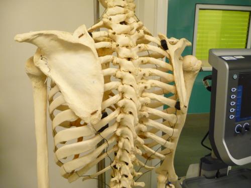
Ultrasound Guided Thoracic Epidural
There are twelve thoracic vertebrae in humans.
The spinous processes of thoracic vertebrae are inclined steepely in
the
upper and mid-thoracic region such that they almost overlap each
other. The tip of the spinous process of the vertebra above lies over
the vertebra below. In the lower thoracic region the inclination is
less steep.
This makes doing thoracic epidural by midline approach very difficult
as the tuohy needle needs to be angulated extremely cephalad and
negotiated through a smaller space between two spinous processes.


The best way to overcome this is by doing a thoracic epidural by
the paramedian
approach especially in high and midthoracic region.
In this approach
once the intervertebral space for doing the epidural is identified, the
tip of the spinous process of the vertebra above is felt and a point
marked one centimeter lateral to it.
Local anaesthetic is injected
to the skin and towards the lamina below. The tuohy needle is inserted
at this point directed perpendicular to the skin to hit the lamina.
This will be the lamina of the vertebra below. Once this is done
the needle is slightly withdrawn and angled medially to so that the
needle walks off the lamina
towards the ligamentum flavum and then the epidural space.
Loss of
resistace to saline or air is used to identify the epidural space.

How can we use ultrasound for thoracic epidural?
To understand this we must first see how the thoracic vertebra looks under
ultrasound and how it is different to lumbar vertebra.

The following video shows what can be seen when we scan over a thoracic
vertebra. Initially the probe is parallel to the long axis of the
spine and then the probe is turned through 90 degrees so that the probe
is perpendicular to the long axis of the spine
Video.
Ultrasound can be used in adults to assist us with thoracic epidural.
With ultrasound we can find out:
1. Midline
2. How deep the lamina is from the skin
3. In some thin adults how deep the dura is from the skin
Once this is determined we can do epidural using paramedian approach as
described earlier without ultrasound. following is a presentation by Dr
Sardesai on use of ultrasound to do lumbar and thoracic epidural
https://1drv.ms/p/s!Aj1UiYRIfQtZgfl6bOk_V34pkHG42A?e=IcMDGd


